
Published on: 18 September 2026
Adults typically have 32 permanent teeth, while children have 20 primary (baby) teeth. In Singapore, dentists use the standardised FDI (Fédération Dentaire Internationale) World Dental Federation notation to identify each tooth precisely. Your teeth are classified into four primary types: incisors (biting and cutting), canines (tearing and jaw guidance), premolars (tearing and grinding), and molars (crushing and heavy chewing, including wisdom teeth).
Understanding tooth names and dental anatomy, including the outer enamel, underlying dentin, and inner pulp, helps you communicate more effectively with your dental practitioner and understand proposed treatment plans.
The Four Main Tooth Types
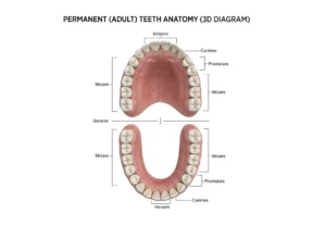
Incisors: 8 front teeth. The four upper and four lower incisors have flat cutting edges and a single root, and perform the first bite of food. Sitting at the front of the arch with a thinner structure, they are the teeth most likely to chip from trauma and the first to show enamel wear from acid erosion or nighttime grinding (bruxism). Exposed dentin after a chip can cause temperature sensitivity and should be checked by a dentist.
Canines: 4 corner teeth. Positioned at the corners of the arch with a single long root, canines have a pointed cusp for tearing food. They also provide “canine guidance”, helping the back teeth disclude during side to side jaw movement and protecting molars from excess lateral force. Canines can occasionally fail to erupt fully and become impacted, particularly in the upper jaw, so their position matters to a balanced bite.
Premolars: 8 middle teeth. Sitting between the canines and molars, premolars usually have two cusps and bridge tearing and grinding. Their pits and fissures trap plaque easily, making them prone to cavities, and in orthodontic treatment they are sometimes selected for strategic extraction to relieve severe crowding.
Molars and wisdom teeth: 12 back teeth. Molars are the largest teeth, with broad, multi-cusped surfaces for final grinding. Clinical studies measuring maximum voluntary biting force confirm that force rises sharply from front to back of the mouth, with molars generating several times the force of incisors. Each quadrant has a first, second and third molar (wisdom tooth), and their deep pits make them prone to decay when hygiene is difficult to maintain.
Why do many adults need their wisdom teeth removed? Third molars usually try to erupt between the ages of 17 and 21, but many jaws lack enough space for them to come through fully. A 2024 systematic review and meta-analysis of impacted third molars worldwide found that Asian populations have one of the highest rates, at 43.1 percent, against a global pooled average of 36.9 percent. A partially erupted or impacted wisdom tooth creates a hard to clean pocket prone to gum inflammation (pericoronitis), decay on the neighbouring molar and localised pain, which is why surgical removal is often recommended.
Primary (Baby) Teeth: 20 Teeth, No Premolars
Children’s 20 primary teeth are made up of incisors, canines and molars only; there are no primary premolars, as permanent premolars later replace the primary molars as the jaw grows. Primary teeth hold space for their successors and support chewing and speech, so they are worth restoring rather than extracting early, since losing one ahead of schedule can let neighbouring teeth drift and crowd out the permanent tooth waiting beneath.
Tooth Numbering System: Understanding Your Dentist’s Shorthand
Singapore dentists use the FDI World Dental Federation two digit notation system to record findings unambiguously. The first digit identifies the quadrant; the second identifies the tooth’s position counting back from the midline.
| Digit | Permanent Quadrant | Digit | Primary (Baby) Quadrant |
|---|---|---|---|
| 1 | Upper Right | 5 | Upper Right Primary |
| 2 | Upper Left | 6 | Upper Left Primary |
| 3 | Lower Left | 7 | Lower Left Primary |
| 4 | Lower Right | 8 | Lower Right Primary |
Second digit: 1 = central incisor, 2 = lateral incisor, 3 = canine, 4 = first premolar (or first primary molar), 5 = second premolar (or second primary molar), 6 = first molar, 7 = second molar, 8 = third molar (wisdom tooth). So tooth 16 is the upper right first permanent molar, and tooth 51 is the upper right primary central incisor. Baby teeth are sometimes recorded with letters (A through T) instead, to keep primary and permanent dentition visually distinct on a chart.
Why do dentists use numbers instead of tooth names? A number removes ambiguity during documentation and referrals. Saying “tooth 46” gives an exact location, whereas “lower right molar” could describe more than one tooth.
Tooth Anatomy: What’s Inside a Tooth
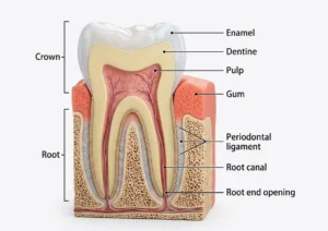
A tooth has two main zones: the crown, the visible portion above the gumline that bears the load of chewing, and the root, usually one to three per tooth, which anchors it into the jawbone below the gum. Within these zones sit three tissue layers.
Enamel, the hard outer layer covering the crown, is the hardest substance in the human body. It has no living cells, so once lost to decay, fracture or acid erosion, it cannot regenerate on its own. Beneath it (and covering the root as cementum) lies dentin, a softer layer with microscopic tubules connecting to the tooth’s nerve; once dentin is exposed, those tubules let cold, heat or touch reach the nerve, which is why it is often sensitive. At the centre sits the pulp, holding the nerve and blood supply. When bacteria reach the pulp and cause irreversible inflammation (pulpitis), this is often the source of a persistent, throbbing toothache, and root canal treatment is usually needed to save the tooth. Left untreated, the infection can progress to a dental abscess and, in more serious cases, spread beyond the tooth, so prompt treatment matters.
Can damaged enamel repair itself? Not structurally. Enamel has no living cells to regrow lost tissue, so a chip, crack or cavity needs a filling, dental crown or bridge, or other restoration. What fluoride and good oral hygiene can do is remineralise very early, microscopic mineral loss on the enamel surface before a cavity forms, which is why early check ups catch problems while they are still reversible.
From Baby Teeth to Permanent Teeth: The Eruption Timeline
The first primary teeth, usually the lower central incisors, erupt around 6 months of age, with all 20 typically present by age 2.5 to 3. Shedding begins around age 6, when permanent incisors and the first permanent molars (the “six year molars”) come in behind the baby teeth, and the transition continues until about age 12 to 13, when the last primary molars are replaced by permanent premolars.
Take Control of Your Dental Health
If you have questions about tooth alignment, wisdom teeth, or routine oral maintenance, a comprehensive clinical evaluation with an examination and diagnostic radiographs can assess the health of your dental structures. Contact us to schedule a gentle oral health assessment with our team.